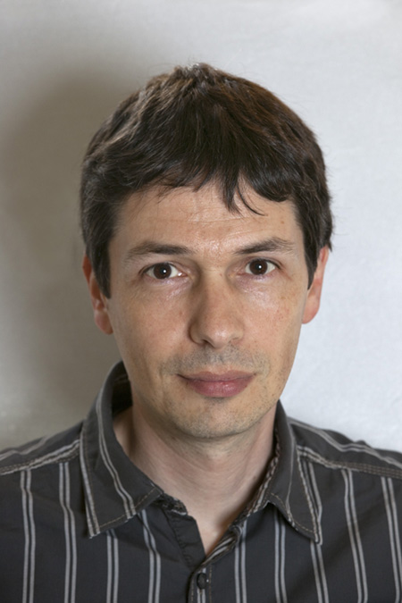
 François Berthod, Ph.D.
Researcher,
Centre de Recherche en Organogénèse Expérimentale de l’Université Laval/ LOEX
CHU de Québec Research Center-Université Laval
Full Professor,
Department of surgery within the Faculty of Medicine of Université Laval
List of publications (external link)
After completing a PhD in Biomedical Engineering from the Université Lyon 1 in France, at the end of 10 years of graduate studies for which I keep an excellent memory, I started, in October 1993, a postdoctoral fellowship at the LOEX under the supervision of Dr. François. A. Auger. Surviving the winter of 93-94, which was the coldest in Quebec in the last 150 years, I set up a reconstructed skin model, based on a collagen-chitosan sponge developed during my Ph.D., in order to graft it on a humanized mouse. In parallel, I have actively contributed to the development of an endothelialized reconstructed skin project initiated by Dr. Auger, for which we have obtained funding for since 1996.
It was in 2000 that I got my first grant from, what was still at the time, the MRC (just before its transfer to CIHR). Also, during this year, I obtained a Junior 1 FRSQ scholarship, the sesame for an official position of Assistant Professor at Université Laval. Although I had to fight hard for subsidies in order to eventually become Full Professor in 2009, it was well worth it, since it is one of the best career in the world! I would like to thank all of my former and current students and research assistants, without whom this entire adventure would not have been possible, for all these exciting projects that we have done together, and all those, even more thrilling, which are looming over the horizon!
My team's research projects are focused on modeling and repairing the nervous system by tissue engineering. We first analyzed the ability of our reconstructed tissue-engineered model to promote effective sensory nerve regeneration in order to recover the tactile sensitivity of the graft after transplantation in mice. We have also shown that the incorporation of laminin, Schwann cells, and immature hair follicles in the reconstructed skin could greatly improve the speed of recovery and quality of the tactile perception. We then produced a model of innervated reconstructed skin to study the influence of sensory innervation on various aspects of the cutaneous physiology, such as angiogenesis. Additionally, we could mimic in vitro several skin pathologies such as diabetic ulcers and psoriasis using cells from taken from patients in order to assess the role of sensory nerves in these diseases.
We have also established a model that allows us to grow motor neurons in three dimensions and to promote the migration of axons that can by myelinated by Schwann cells. These neurons can be combined with astrocytes and microglia to mimic the spinal cord. This model is being developed to mimic in vitro the process of motor neurons degeneration in amyotrophic lateral sclerosis (ALS). Cells from mice engineered to develop the disease as well as cells from patients can be used for that purpose.
We have also developed an endothelialized nervous tube produced by tissue engineering to repair transected peripheral nerves. It was grafted on immunodeficient rats to assess its ability to promote hind motor recovery after implantation within the sciatic nerve.
The main purpose of my research is to use tissue engineering techniques of in vitro organ reconstruction to study different diseases that can be induced or modulated by the nervous system to better understand their mechanism and find new therapeutic approaches.
We have produced a model of innervated reconstructed skin to investigate the influence of various parameters on sensory innervation of skin physiology, such as angiogenesis. We then developed skin models to recapitulate in vitro the phenotypes of skin pathologies such as diabetic ulcers and psoriasis, using cells from patients in order to assess the role of sensory nerves in these diseases. The effect of sensory innervation can be studied in vitro in our models and in vivo after their transplantation in mice.
We have also established a model to grow motor neurons in a three dimensional enviroment and to promote the migration of axons while achieving their myelination by Schwann cells. These neurons can then be combined with astrocyte and microglia to mimic the spinal cord. This model is being developed to reproduce the in vitro process of motor neurons degeneration in amyotrophic lateral sclerosis, using cells derived from mice developing the disease, or ALS patients cells.
The peripheral nerve transection induced by a traumatic, iatrogenic or neoplastic origin causes major sensory and motor impairments that may lead to a complete paralysis of a limb (hand, arm, leg), which is very debilitating for the patient. Our goal is to develop a tissue-engineered nerve tube reconstructed from the patient's own cells, and which can be grafted between the two stumps of the severed nerve to allow nerve regeneration. Moreover, our approach aims to fill high caliber gaps over great distances. For that purpose, we will incorporate in our preformed tube a network of capillaries that will induce rapid vascularization of the implant to the internal nerve vascular network after transplantation. We will also incorporate into the tube, in collaboration with Dr. Hélène Khuong, a clinical investigator and Neurosurgeon at the CHU de Québec-Université Laval, autologous Schwann cells to enhance axonal migration into the graft. The diameter and length of the tube may be adjusted to the characteristics of the nerve to repair, and may greatly improve motor function recovery of patients.
The main purpose of my research is to use tissue engineering techniques of in vitro organ reconstruction to study different diseases that can be induced or modulated by the nervous system to better understand their mechanism and find new therapeutic approaches.
We have produced a model of innervated reconstructed skin to investigate the influence of various parameters on sensory innervation of skin physiology, such as angiogenesis. We then developed skin models to recapitulate in vitro the phenotypes of skin pathologies such as diabetic ulcers and psoriasis, using cells from patients in order to assess the role of sensory nerves in these diseases. The effect of sensory innervation can be studied in vitro in our models and in vivo after their transplantation in mice.
